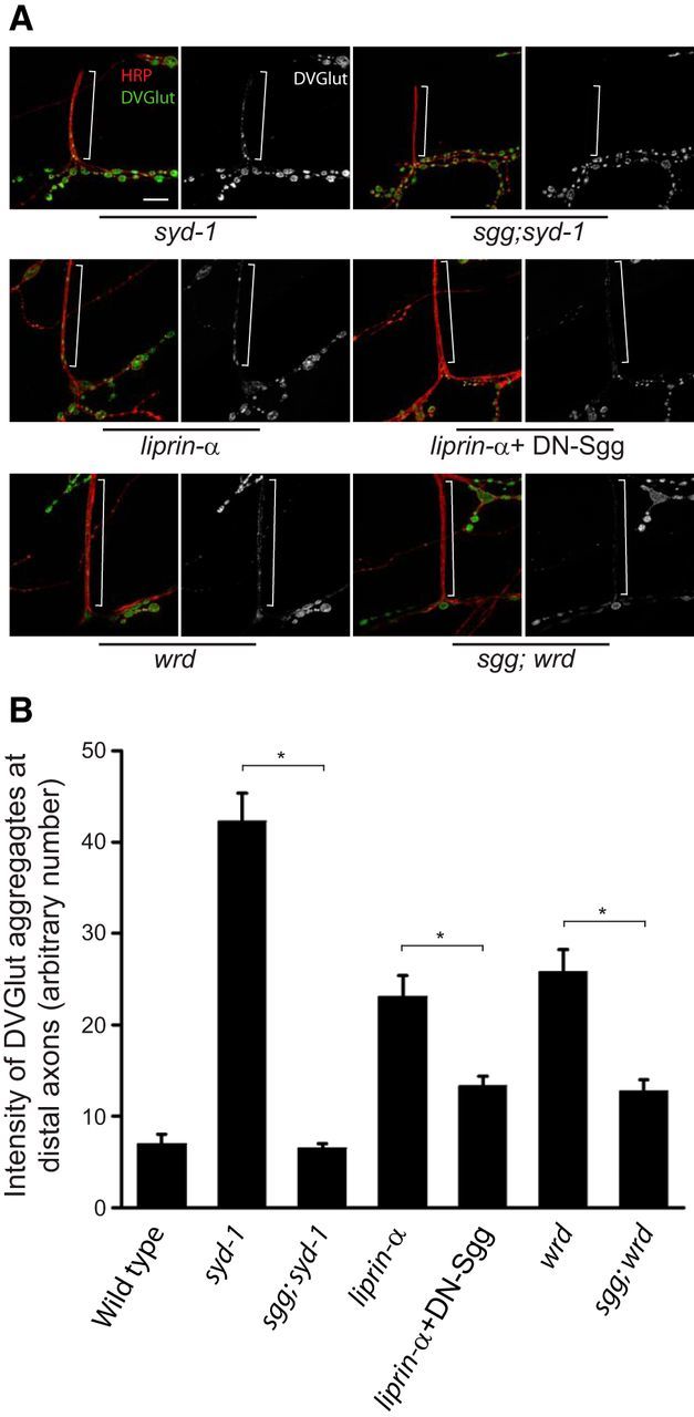
Figure 5.

Loss of GSK-3β function suppresses the syd-1, liprin-α, and wrd mutant distal axon phenotype. A, Representative confocal images of the distal axons of muscle 12/13 NMJs costained with DVGlut and HRP in third-instar larvae of syd-1 (syd-1ex1.2/syd-1ex3.4), sgg;syd-1 (sggG0055;syd-1ex1.2/syd-1ex3.4), liprin-α (liprin-αEPexR60/liprin-αF3ex15), liprin-α + DN–sgg (BG380–Gal4;liprin-αEPexR60/liprin-αF3ex15,UAS–sggA81T), wrd (wrd104/wrd104), and sggG0055;wrd (sggG0055;wrd104/wrd104) mutants. The reason for using the dominant-negative UAS–Sgg transgene instead of the sgg mutant with liprin-α mutant is that the second-chromosome-balanced sgg mutants are lethal. White brackets show terminal axons crossing muscle 13 to innervate muscle 12. Scale bar, 10 μm. B, Quantification of the distal axon vesicle accumulation (measured by average intensity of DVGlut at muscle 12/13 terminal axons) in third-instar larvae of wild-type and flies presented in A. n = 6, 7, 12, 12, 16, 11, and 8, respectively. *p < 0.001. Error bars denote SEM.
