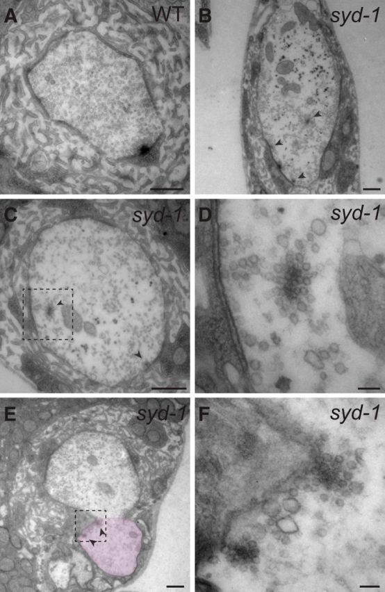
Figure 6.

EM analysis of the ectopic vesicle accumulation. A–D, Electron micrographs of wild-type (WT) and syd-1 (syd-1ex1.2/syd-1ex3.4) mutant NMJ boutons. E, F, Electron micrographs of syd-1, a mutant NMJ bouton, and its connected axonal tip (pink-marked area in E). Identification of the axonal tip was based on the absence of subsynaptic reticulum (SSR) and the presence of glial wrapping. Arrowheads mark floating high-electron-dense materials surrounded by vesicles in syd-1 mutant samples. Some of the floating high-electron-dense materials are very close to or almost touching the plasma membrane. D and F show high-magnification images of dashed boxes in C and E, respectively. Scale bars: A, B, C, E, 0.5 μm; D, F, 80 nm.
