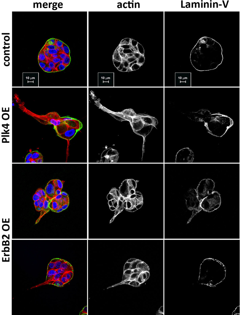
Extended Data Figure 4. Similarity between cells with centrosome amplification and cells with oncogene-induced invasion.
Cells were stained for F-actin (red), laminin-V (green) and DNA (blue). Similarity between the invasive protrusions of cells with extra centrosomes and the ones generated by cells overexpressing ErbB2, as previously reported4,42. In both conditions, invasive protrusions are characterized by the formation of actin-rich protrusions that are accompanied by degradation of the basement membrane. Scale bar: 10µm.

