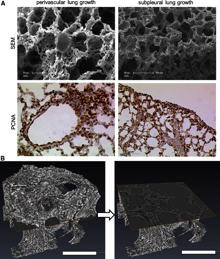
Fig. 2.
Spatial growth pattern of cardiac lobe. a The spatial heterogeneity of different lung growth can be seen in SEM-images of microvascular corrosion casts (above) and anti-PCNA-staining (below): perivascular lung growth located mainly in central parts of the lung (left panel) and subpleural lung growth (right panel) containing numerous anastomoses with pleural vessels. b High resolution synchrotron radiation tomography revealed the compact zones of vascular growth upon cross-section. Bar = 100 μm

