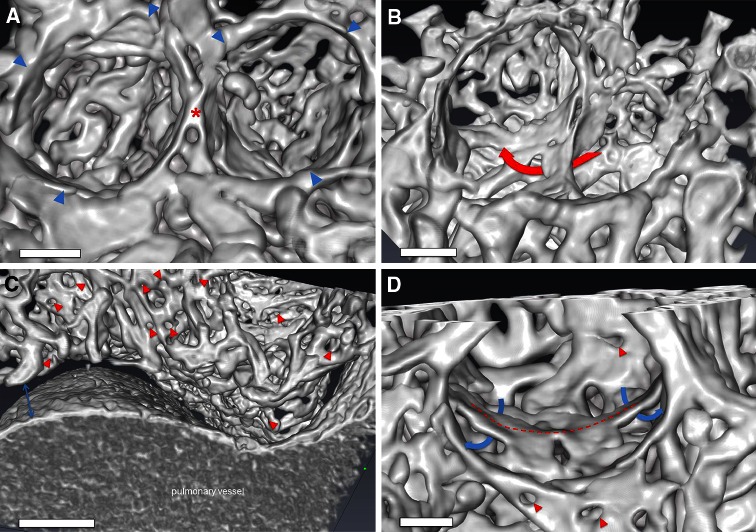
Fig. 6.
Synchrotron radiation tomographic microscopy. a Three-dimensional evaluation of microvascular corrosion casts by SRXTM illustrating the alveolar basket structure accompanied by the limiting AER vessels (blue arrowheads) and the elevated ridge (asterisk). Bar = 15 μm. b A typical example of double-layered vessels (red arrow) during alveolarization. Bar = 20 μm. c Analysis of central areas of the cardiac lobe revealed an increased density of intussusceptive holes (red arrowheads) indicative of the occurrence of intussusceptive angiogenesis around larger vessel structures. Bar = 60 μm d In lung alveolarization a ridge (dashed line) can be seen in the midline of the alveolar basket accompanied by double capillary layers (blue arrows) enabling the lifting-off of the inter-airspace septum. The rapid expansion by intussusceptive angiogenesis (red arrowheads) allows the pacing of isotropic lung growth after pneumonectomy. Bar = 10 μm (see movies in supplemental material)

