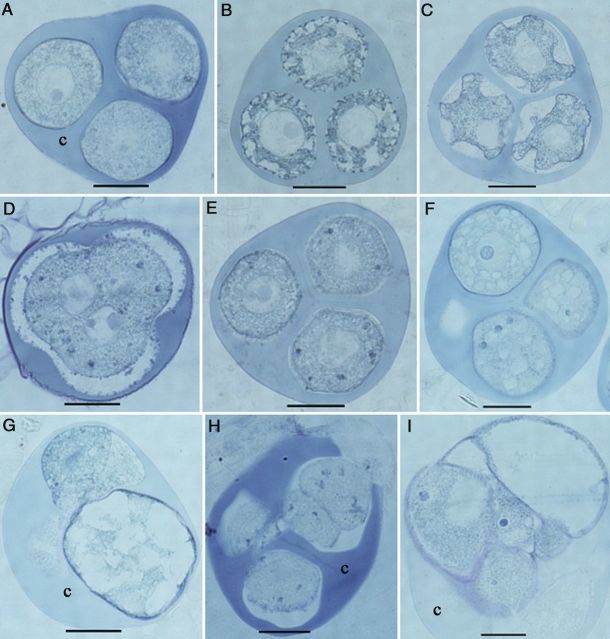
Figure 10.
Histological aspects of the microspore reprogramming of M. esculenta genotype TMS 60444. (A–C) Fresh tetrads. (A) Immature tetrad microspores with smooth surface. (B) Tetrad microspore with initial deposition of exine particles and initial inward movement of cytoplasm. (C) Mature tetrad microspores with deep folds forming on the irregular surface of the microspore. (D) A meiocyte among the cultured tetrads. Note that cytokinesis has not yet occurred even after meiotic karyokineses. (E) Significant appearance of protein deposits after applying 3 days of cold pretreatment (blue particles). (F) Small vacuoles in microspores 1 day after culture. (G) Enlarged microspore with large vacuoles 3 days after culture. (H) Cell division in microspores 3 days after culture. (I) A multi-cellular structure still enclosed in the callose wall (c) 7 days after culture (scale bars = 20 µm).

