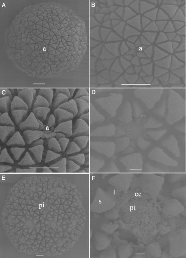
Figure 7.
Scanning electron micrographs showing aspects of cultured tetrads of M. esculenta genotype SM 1219-9. (A) A fresh microspore. Note the differential size and shape of sculptured particles at the aperture (a) area as compared with the rest (scale bar = 10 µm). (B) Close-up view of the pattern of exine sculptures at the aperture of a fresh microspore (scale bar = 10 µm). (C and D) Changes occurring at the aperture in the enlarging microspore. Note that the aperture is opening up and the intine becomes visible (scale bars (C) = 10 µm and (D) = 2 µm). (E) A microspore with protruded intine (pi) through the aperture (scale bar = 10 µm). (F) Close-up view of an aperture where the exine particles are much expanded, the aperture has opened well and where the intine is coming out, making a protrusion. Note the different layers of the exine: end exine (ee), tectum (t) and the sculpture particles (s) located on the tectum (scale bar = 2 µm).

