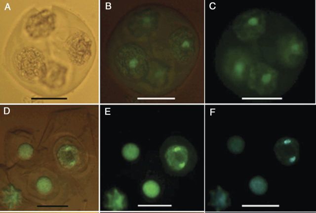
Figure 8.
Fluorescence micrographs of nuclei in cultured tetrads of M. esculenta genotype TMS 60444. (A–C) A fresh tetrad without fluorescence and with partial and full fluorescence under a green filter. (D–F) Cultured tetrad containing an enlarged microspore undergoing mitosis under partial and full fluorescence with a green filter and full fluorescence with a blue filter (scale bars = 22 µm).

