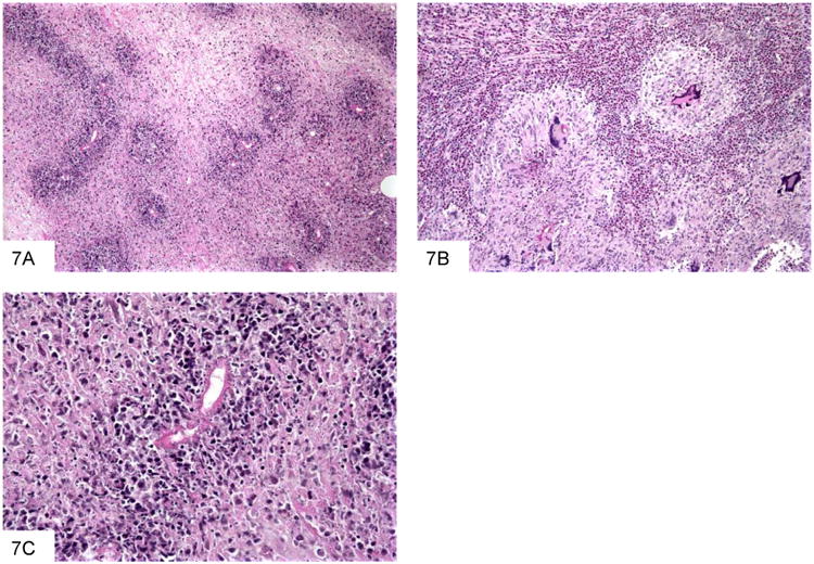
Figure 7.

This section of a paracolonic mass due to B. ranarum shows extensive necrosis and scattered organisms highlighted by the bright pink protein deposition known as Splendore-Hoeppli phenomenon (A). Basidiobolomycosis often features granulomas with giant cells, and abundant eosinophils (B). This high power view shows a fungus outlined by bright pink proteinaceousmaterial (C).
