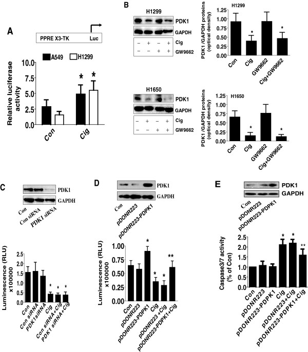
Figure 2.

Ciglitazone inhibited PDK1 protein expression independent of PPARγ. A, H1299 and H1650 cells were transfected with control or PPRE X3-TK-luc reporter (from Addgene) for 24 h, followed by treating with ciglitazone for an additional 24 h. Afterwards, the Luciferase reporter activity was measured using Luciferase Assay System (Promega) according to manufacturer's instructions. The bars represent the mean ± SD of at least three independent experiments for each condition. *indicates significant difference as compared to the untreated control group (P < 0.05). B, Cellular protein was isolated from H1299 and H1650 cells cultured for 1 h in the presence or absence of GW9662 (20 μM) before exposing the cells to ciglitazone (20 μM) for an additional 24 h, then subjected to Western blot analysis. C, H1299 cells were transfected with control or PDK1 siRNA (80 nM) for 40 h, followed by exposing the cells to ciglitazone (20 μM) for an additional 24 h. Afterwards, the luminescence of viable cells was detected using Cell Titer-Glo Luminescent Cell Viability Assay kit. D-E, H1299 cells were transfected with the control and PDK1 expression vectors using the oligofectamine reagent according to the manufacturer’s instructions. After 24 h of incubation, cells were treated with or without ciglitazone for an additional 24 h. Afterwards, the luminescence of viable cells was detected using Cell Titer-Glo Luminescent Cell Viability Assay kit (D). In separate experiment, the relative caspase 3/7 activity (E) is indicated as percentage of untreated control cells. The bars represent the mean ± SD of at least four independent experiments for each condition. Insert on the top panel shows a Western blot for PDK1 protein. *indicates significant difference as compared to the untreated control group (P < 0.05). **Indicates significance of combination treatment as compared with ciglitazone alone (P < 0.05).
