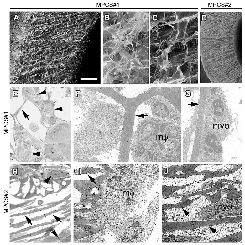
Fig. 1. Scaffolds with radially oriented porosity.
Scanning electron micrographs of MPCS#1 (A–C) and MPCS#2 (D). MPCS#1 with a gradient in radially oriented pore distribution of the wall (A): smaller porosity of the external portion (B) compared to internal portion (C); MPCS#2 with uniform and smaller pore distribution (D). Semithin sections and electron microscopy of MPCS#1 (E–G) and MPCS#2 (H–J)-implanted rats after 8 days. In MPCS#1, macrophages (E, arrowhead; F, mf) and myofibroblast (G, myo) between the pores and attached to the collagen micro-structure (E–G, arrows). In MPCS#2 few macrophages (H, arrowhead and I, μm) and myofibroblasts (J, myo) are present (H–J, arrows: collagen micro-structure). Scale bar: in A and D 100 μm; 20 μm for B, C, E and H; 2 μm for F, G, I and J.

