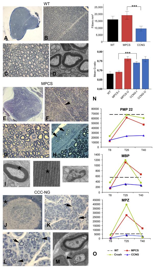
Fig. 5. Nerve regeneration.
Morphological analysis of WT sciatic nerve (A–D), MPCS (E–I) and CCNG (J-M)-implanted rats after 60 days. Semithin cross section of entire normal sciatic nerve (A): fiber density, myelinatyon and endoneurium are normal (BeD). In MPCS group, scaffold is completely substituted by normal nerve (E) with normal amount of myelinated fibers (F and G); the stromal tissues is re-established with intrafascicular septum (F, arrowhead). In the perineurium, clusters of regeneration are present (H, arrow); electron microscopy shows compacted myelin (I, asterisk) and normal non-myelinated fibers (I). In CCNG group the scaffold wall is still present (J, asterisk), the endoneurium is characterized by minifascicle formations (K, arrowhead) with thin myelinated fibers grouped in clusters of regeneration (L and M, arrows). N: fiber density and g-ratio in sciatic nerve from WT, MPCS and CCNG inside the scaffold (−I) and outside (−O) at tibial nerve (***= p < 0.0005). O: GE pattern of key protein component of myelin in PNS. Scale bar: 100 μm A, E and J; 40 μm B, F and K; 20 μm C, G, H, L, N, O and P; 2 μm D, I and M.

