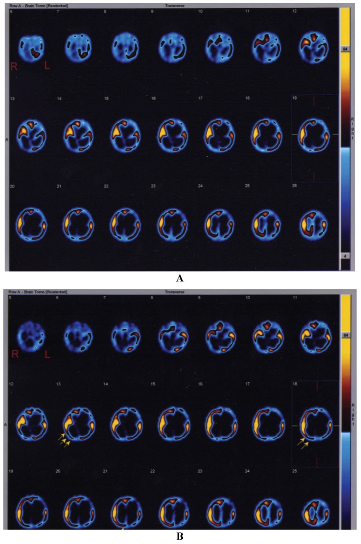
Figure 1.
Single-photon emission computed tomography (SPECT) study showing the brain perfusion pattern in the second infant affected by severe hypoxic-ischemic injury. (A) Before treatment. (B) After intraventricular NGF infusion. The arrows in (B) highlight the sites of improvement of cerebral perfusion in right temporal and occipital cortices.

