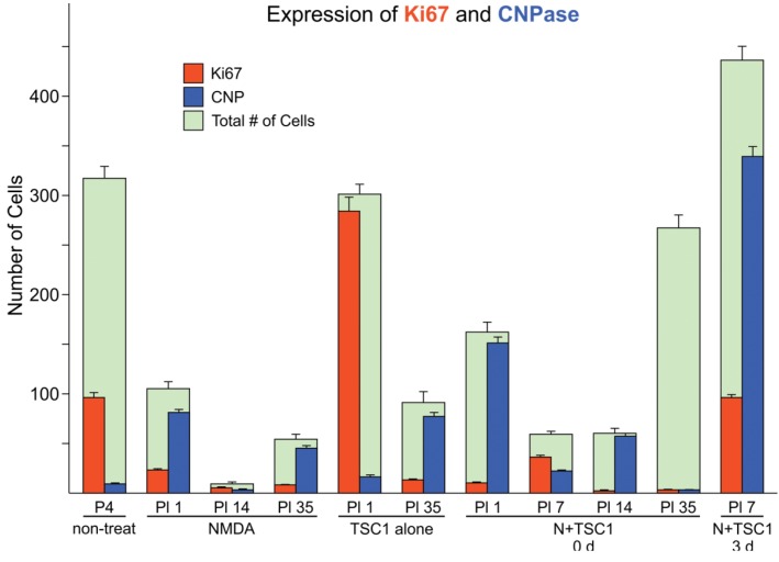
Figure 2.
Cell loss in the subventricular zone (SVZ) is partially rescued in the presence of TSC1 via cell survival or cell proliferation. Double immunofluorescence: cells expressing the proliferation marker Ki67 were found in this region at early time points in the presence of TSC1. Some cells co-expressed the two markers (Ki67/CNPase). In contrast, when N-methyl-d-aspartate (NMDA) was injected alone there was a dramatic reduction of the total number of cells. Green bars represent the total number of cells (i.e., 100% or the total number of cells counted in that field. Numbers for saline and non-treated mice were very close with no significant differences. Values are expressed as mean ± SEM of the counts of 9 fields per area from three independent experiments. p < 0.05 vs. controls. All differences with respect to non-treated mouse brains as well as, across treatments were significant. P5 = Postnatal day five; PI = post injection day.

