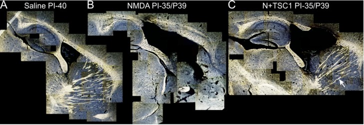
Figure 4.
Myelination is partially rescued from excitotoxicity by TSC1. Representative para-sagittal brain sections (28 μm thick) stained with the Spielmeyer’s method for frozen sections. These views show (A) the myelination pattern with saline treatment 40 days post-injection (PI). The extent of tissue damage, ventricle enlargement, and myelin loss in mice treated with NMDA (B) and its recovery with TSC1 (C) treatment. Moreover, NMDA treated mice showed areas where tissue was spared in the CC and CPu but there was not myelin staining. In contrast, mice treated with TSC1 showed nice myelinated fibers indicating that after the excitotoxic insult functional OLs developed and actively myelinated axons. The arrow points to a bundle of myelinated axons in the CPu of a mouse treated with NMDA + TSC1 simultaneously. In contrast, the same region of mice injected with NMDA alone did not show myelinated axons. The variability within each treatment group, consisting of 6 animals, was minimal based on low magnification examination as shown in Figure 4.

