
Figure 13.
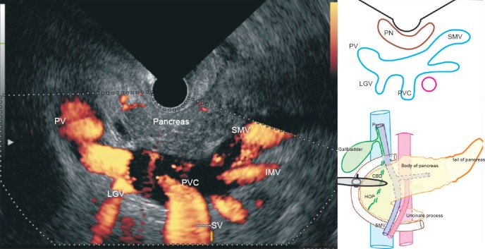
The endosonographic image, in a case of portal hypertension from the duodenal bulb, shows the normal anatomy of the main portal vein at its formation. The superior mesenteric vein is seen merging with the splenic vein to form the portal vein. The left gastric vein is seen joining the upper border of the portal venous confluence. Inferior mesenteric vein is seen joining the lower border of the portal venous confluence.