
Figure 6.
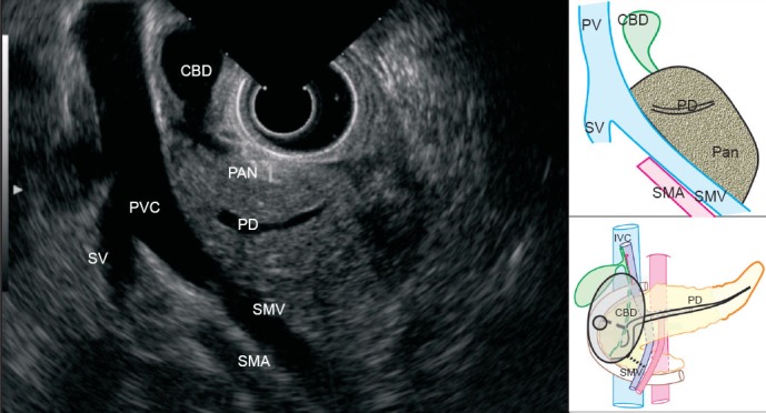
The scope is pushed in the duodenal bulb and the balloon is inflated. In this position, the main portal vein is seen in the longitudinal section. The superior mesenteric vein is seen joining the splenic vein to form the main portal vein behind the common bile duct. The pancreatic duct is also seen from the 4 o’clock to the 7 o’clock position. The portal vein is radiologically up to 1.6 cm in diameter and about 8 cm long.