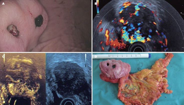
Figure 6.

Gastrointestinal stromal tumor. A: Typical endoscopic features; B: CEHMI-EUS image of the same tumor, and please notice the rich vessel system; C: CELMI-EUS image, and the necrotic area was displayed in the upper area of the tumor; D: The surgical specimen of the tumor. CELMI-EUS: contrast-enhanced low-MI endoscopic ultrasound; CEHMI-EUS: contrast-enhanced endoscopic Doppler ultrasound with high-mechanical index.
