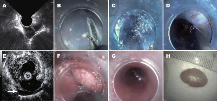
Figure 1.

The tunnel type endoscopic submucosal dissection procedure and EUS in tunnel. A: Muscle layer tumor viewed on EUS; B: Longitudinal mucosal incision; C: Submucosal tunnel creation; D: Endoscopic view of tumor in tunnel, difficult to distinguish from the muscular layer or aortas; E: Tumor(T) identified by EUS in tunnel. A small amount of irrigated saline leaked through the muscular layer of esophagus to the mediastinal space (arrow); F: Tumor dissection in the tunnel; G: Mucosal entrance closing; H: Tumor ex vivo. EUS: endoscopic ultrasound. T: tumor.
