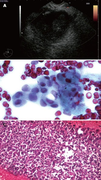
Figure 4.

Endoscopic ultrasound-guided fine needle aspiration performed with a 22-G needle on the side of a hypoechoic pancreatic tumor mass, with both cytology and microhistology performed after at least 3 passes. Cytology showed a clump of malignant cells, while microhistology confirmed the diagnosis of adenocarcinoma.
