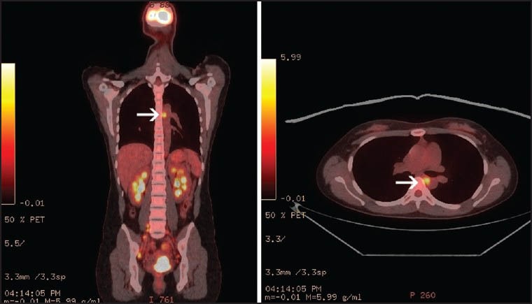
Figure 1.

Positron emission tomography-computed tomography scan demonstrated hypermetabolic primary cervical mass. A softtissue density in the posterior mediastinum with standard uptake value of 5.2 presumed as paraesophageal posterior mediastinal lymphadenopathy (white arrow)
