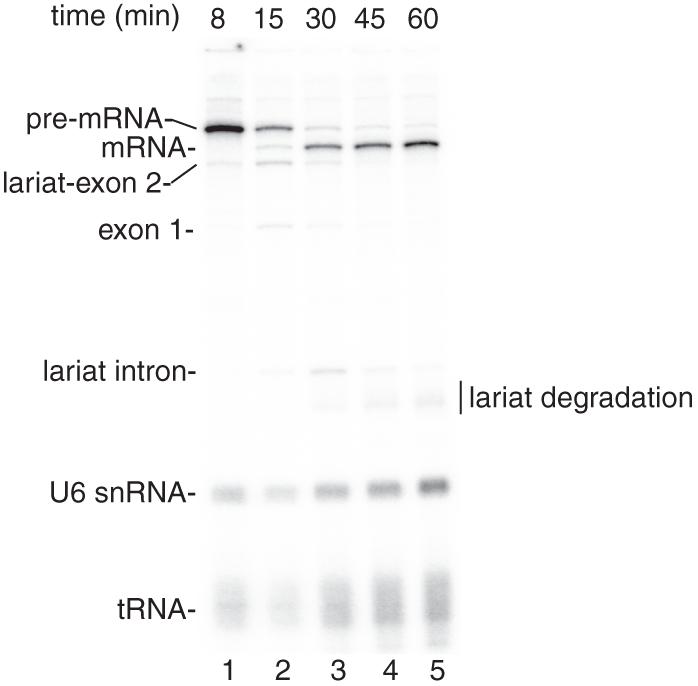
Figure 1.
Coupled RNAP II transcription and pre-mRNA splicing in vitro. (A) Structure of the DoF CMV-DNA template. The sizes of the exons and intron are indicated. B. 32P-UTP and the CMV-DoF DNA template were incubated under transcription/splicing conditions for 8 minutes. α-amanitin was added after the 8-minute time point and incubation was continued for the indicated times. Pre-mRNA and the splicing intermediates are indicated.The endogenous U6 snRNA and tRNA in the extract are labeled by the 32P-UTP (see Reddy et al. [1987].

