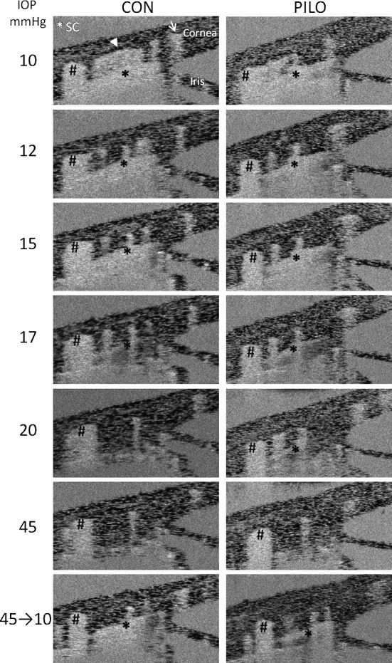
Figure 5.

Effects of sequential IOP steps on speckle variance in anterior eye of mouse in the absence (CON) or presence of pilocarpine (PILO). Shown are speckle variance OCT equivalents of the averaged images of iridio-corneo angle tissues containing SC lumen (asterisk) in Figure 3. Left column of images displays effect of sequential IOP steps on speckle variance in SC lumen from the same sagittal section. Note that as IOP increases, speckle variance in SC decreases, as well as does downstream scleral vessels (arrowhead), but not neighboring scleral vessels (hashmarks and arrow). The effects of pilocarpine on SC speckle variance from the same sagittal section of the same mouse at sequential IOPs are shown in the right column of images alongside untreated. Shown are representative data from one mouse of five total mice that were examined.
