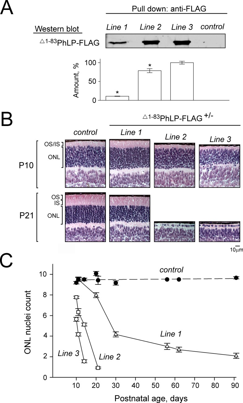
Figure 1.
Suppressing the CCT activity in mouse photoreceptors by phosducin-like protein short. (A) Whole-retina extract from 8-day-old mice were analyzed by pull down with anti-FLAG agarose. A representative Western blot shows the amounts of captured Δ1-83PhLP-FLAG, as visualized with antibody against FLAG, in transgene-positive (Δ1-83PhLP-FLAG±) and control (Δ1-83PhLP-FLAG−/−) mice from the lines 1 to 3. Graph: protein bands in each experiment were quantified and their values expressed as percent of the highest value found in line 3. Bars are SEM with n = 3, and P < 0.05 as determined by paired t-test. (B) Paraffin-embedded retina cross-sections were stained with hematoxylin and eosin to visualize its cellular composition at the age P10 and P21. (C) Nuclei count across the outer nuclear layer (ONL) as a function of mouse age. Bars are SEM, n = 6. OS, outer segments; IS, inner segments.

