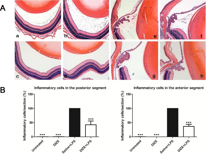
Figure 3.
Histological evaluation of EIU from hematoxylin and eosin–stained paraffin sections with and without systemic DIZE treatment. (A) Representative images showing infiltrating inflammatory cells in the posterior and anterior segments of the eyes in the DIZE + LPS group (a, e), the saline +LPS group (b, f), untreated group (c, g), and DIZE group (d, h). (B) Quantification of the percentage of inflammatory cells in the posterior and anterior segments of the eyes of all groups. ***P < 0.001 (n = 6, versus saline + LPS group; ×100 magnification for a–h).

