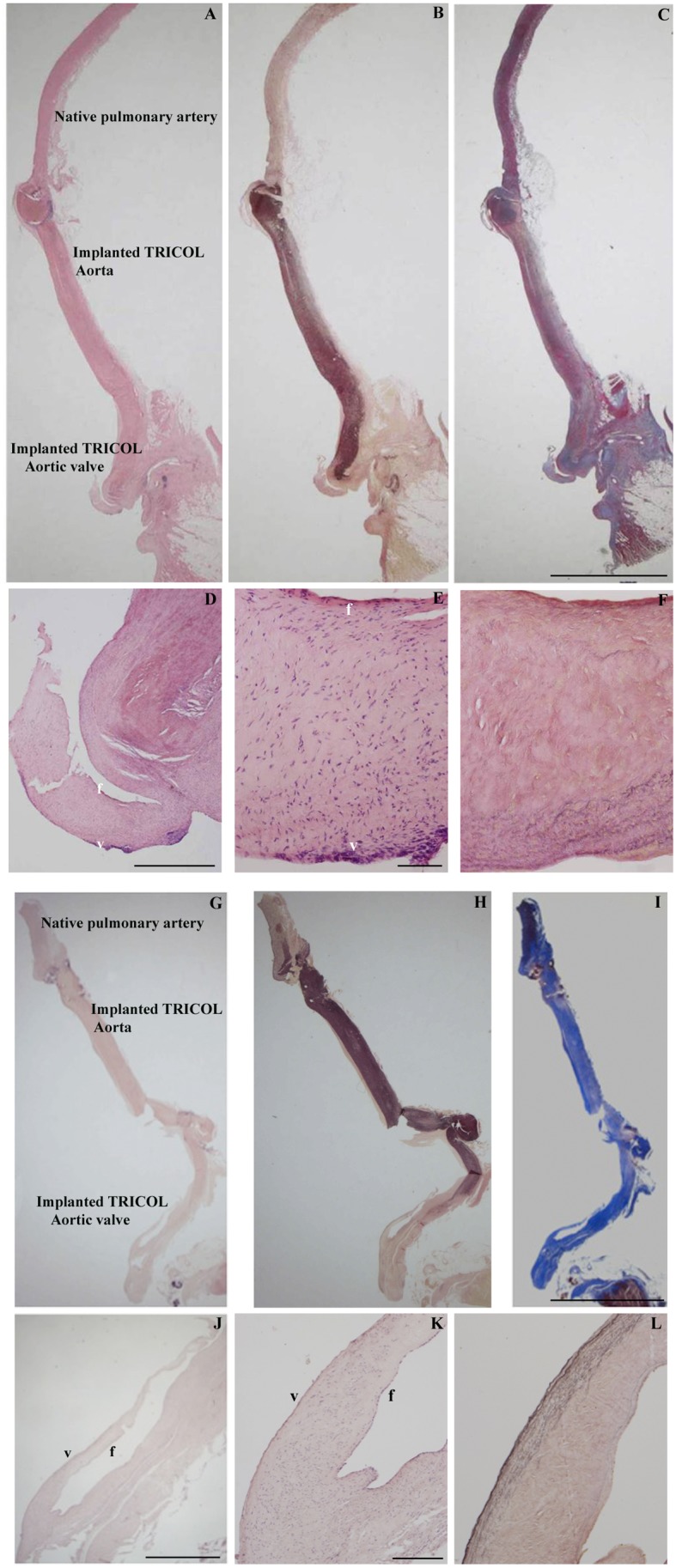
Figure 2. Histologic evaluation of explanted allografts.

Panoramic (A and G: H&E, B and H: Elastic van-Gieson, C and I: Azan’s Heidenhain trichrome, magnification: 5 cm) and close-up (D–F and J–L) allograft views respectively at 6 and 15 months after surgery: note trilaminated arrangement with recipient’s repopulating cells on both leaflet sides, i.e. ventricularis (v) and fibrosa (f) (D–E, J–K: H? F and L: Elastic van-Gieson). Magnifications: (A–C and G–I) 1 cm; (D) 500 µm; (E, F) 100 µm; (J) 700 µm; (K, L) 200 µm.
