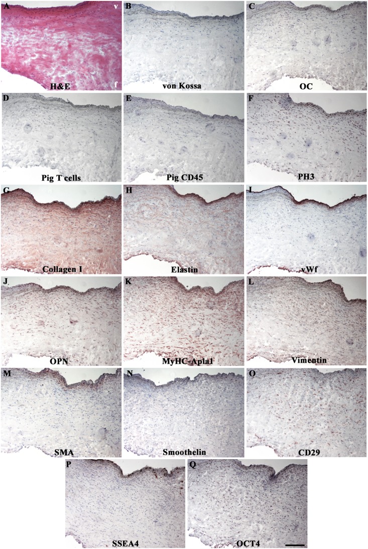
Figure 4. Immunohistochemical profile of recellularized leaflets at 6 months.
Undetectable calcifications (B, C) or immune rejections against allogeneic tissues (D, E). Conserved trilaminated ECM architecture (A, G and H). Native-like EC (I) and VIC phenotypes (J–N). Stem cell markers of mesenchymal (O) and embryonic (P and Q) lineages, mainly expressed in ventricularis. In (F), PH3-positive leaflet-colonizing cells. V = ventricularis; f = fibrosa. Magnification: 200 µm.

