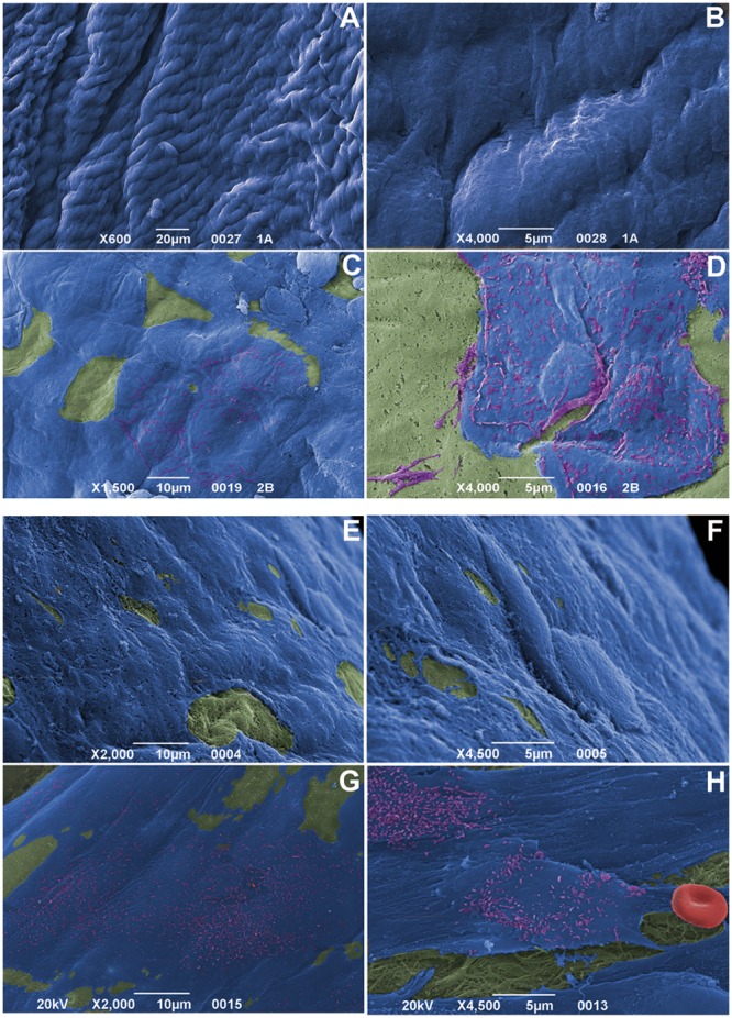
Figure 6. Scanning electron microscopy on allograft cusps at 6 and 15 implantation months.

Almost complete endothelial coverage (blue), progressive EC acquisition of surface microvilli (purple) and no platelet aggregation onto fibrosa (green; at 6 months in A–B, after 15 months in E–F) and ventricularis (green; at 6 months in C–D, after 15 months in G–H). Note the absence of fibrin deposition, as well as no red cell (red) entrapment in H. Magnifications: (A) 20 µm; (B, D, F, H) 5 µm; (C, E, G) 10 µm.
