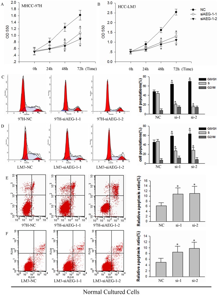
Figure 2. AEG-1 knockdown induces growth inhibition and cell death in MHCC-97H and HCC-LM3 cells.
(A and B) Effects of AEG-1 silencing on cell proliferation were measured by MTT assay in (A) MHCC-97H and (B) HCC-LM3 cells over a time course of transfection (*P<0.05). NC, transfected with a scrambled siRNA as a negative control; siAEG-1-1 and siAEG-1-2, transfected with siRNA targeted against AEG-1. (C and D) Propidium iodide staining and cell cycle analysis was performed by flow cytometry for (C) MHCC-97H cells and (D) HCC-LM3 cells transfected with NC siAEG-1-1 or siAEG-1-2. Samples were assayed 72 h after transfection. The cytometric profile of a representative experiment which containing three (left, middle and right) cures on behalf of cell cycle (G0/G1, S, and G2/M) stage is shown (left). Area of different cures corresponding cell cycle stages in three independent experiments are quantified (right; *P<0.05). (E and F) Cell viability analysis of MHCC-97H and HCC-LM3 cells transfected with siAEG-1-1 and siAEG-1-2 for 72 h was measured. Results from a representative experiment are shown (left) and the mean±SD of three independent experiments (right; *P<0.05).

