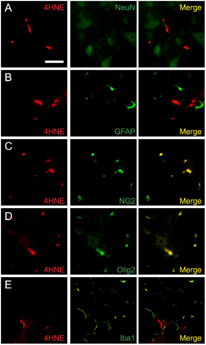
Figure 7. 4-HNE were mainly produced on oligodendrocyte in the spinal cord 4 days after PSNL.
(A–E) Double immunostaining of 4-HNE with cellular markers for neurons (A), astrocyte (B), oligodendrocyte (C, D), and microglia (E). 4-HNE signals were mainly colocalized with the oligodendrocyte marker NG2 and olig2, but not with NeuN, GFAP, and Iba1 in the spinal cord at the end of 4 days period after PSNL. Scale bar: 20 µm.

