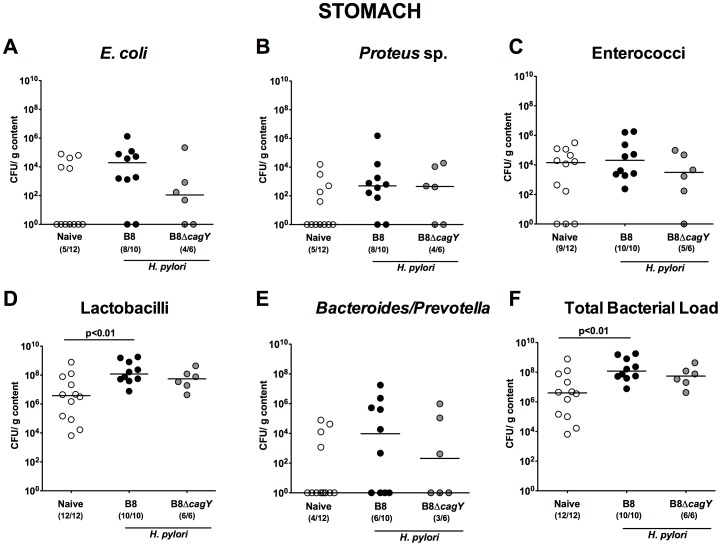
Figure 3. Stomach microbiota composition in Mongolian gerbils 14 months following H. pylori infection.
Fourteen months following oral infection of Mongolian gerbils with H. pylori wildtype strain B8 (B8; black circles) or H. pylori mutant strain lacking cagY (B8ΔcagY; grey circles), the microbiota composition of luminal stomach contents were quantitatively analyzed by culture as described in Methods. Uninfected age-matched animals served as negative controls (Naïve; white circles). Numbers of live (A) E. coli, (B) Proteus sp., (C) Enterococci, (D) Lactobacilli, (E) Bacteroides/Prevotella spp. as well as the (F) total bacterial load are indicated as colony forming units (CFU) per g luminal content. Numbers of animals harboring the respective bacterial species are given in parentheses. Medians and significance levels (p-values) determined by Mann-Whitney-U test are indicated. Data shown were pooled from three independent experiments.

