
Figure 7.
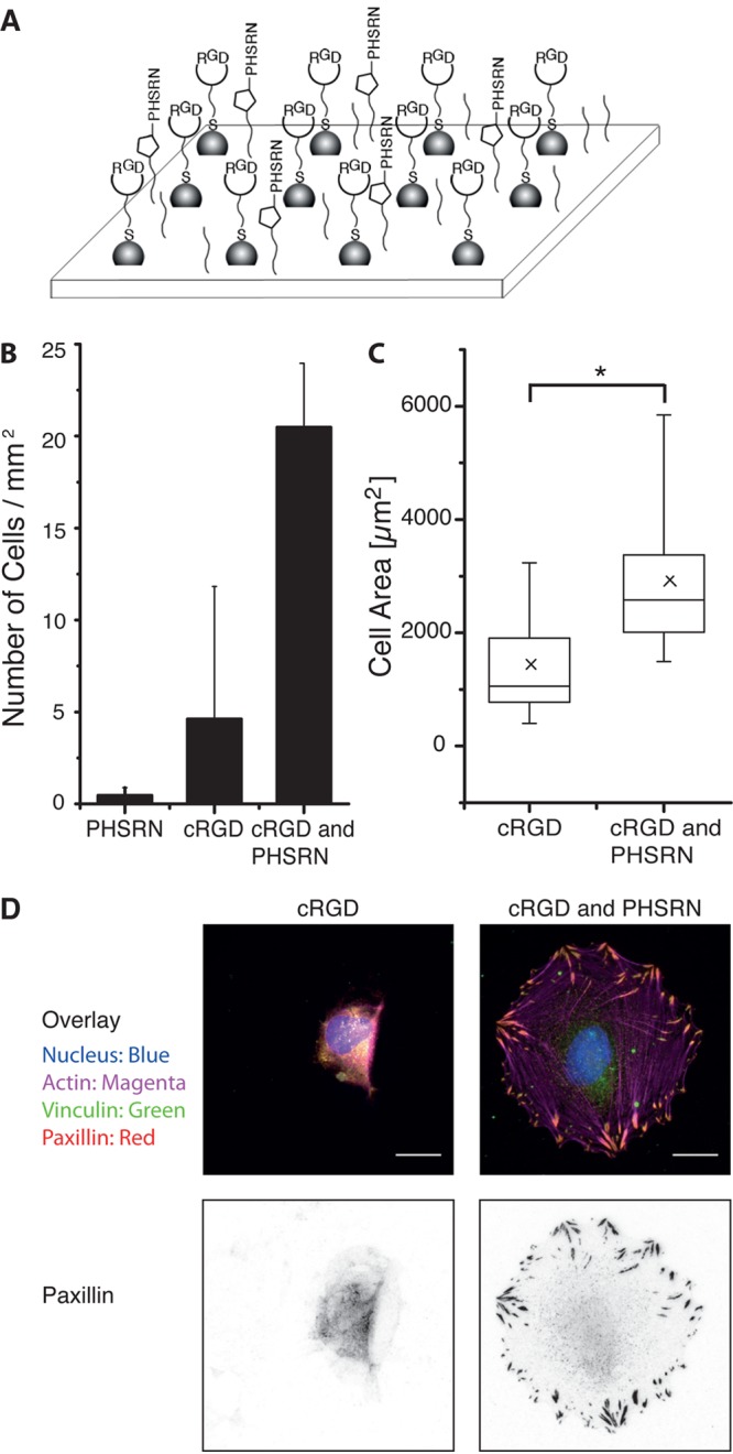
(A) Scheme for the dual functionalization with adhesion peptide cRGD and synergy peptide PHSRN. (B) Density of adherent REF WT cells on substrates with gold nanoparticles and PEG-alkyne (10 mol %) functionalized with adhesion peptide cRGD and/or synergy peptide PHSRN. The average distance of the gold nanoparticles is 100 nm. (C) The spreading area of cells on cRGD- or cRGD- and PHSRN-functionalized surfaces. (*p < 0.0001) (D) Fluorescent images of adherent cells on substrates functionalized with cRGD or cRGD and PHSRN. The nucleus is shown in blue, actin in magenta, vinculin in green, and paxillin in red. The vinculin and paxilin stains colocalize. In particular, the inverted paxilin staining (right) demonstrates the formation of mature focal adhesions on the bifunctional surfaces. The scale bar is 20 μm.