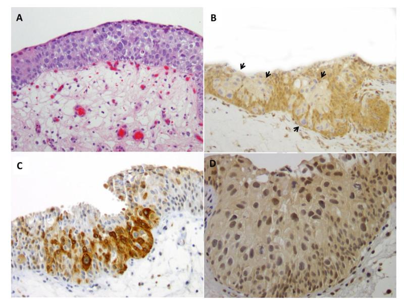
Figure 1.
A (H&E). Urothelial carcinoma in situ composed of large and pleomorphic tumor cells. Mitotic figures present. B (PTEN). Loss of PTEN expression (0 intensity) in tumor cells (arrows) compared to adjacent non-neoplastic urothelium. C (p-S6). Tumor cells with strong expression (3+ intensity) of p-S6 compared to adjacent non-neoplastic urothelium. D (p-Akt). Increased expression of p-Akt (2+ intensity) in tumor cells.

