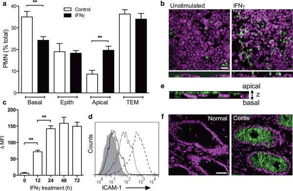
Figure 1. IFNγ-induced expression of ICAM-1 promotes PMN retention on the apical epithelial membrane.
(A) PMN were stimulated (100nM fMLF) to migrate across control or IFNγ treated T84 IECs. Non-migrated PMN (basal), PMN within the epithelial layer (epith), PMN associated with the apical IEC membrane after TEM (apical), and PMN that completed TEM (TEM) were quantified. (B) Following 1h of TEM, IEC monolayers were fixed and stained for cell nuclei (blue) and PMN Mac-1 (green). Representative images show enhanced apical attachment of PMN after migration across IFNγ treated T84 IECs. The bar is 20μm. Bottom panels are projections of images acquired in series in Z-direction. (C-F) Confluent T84 IECs were treated with IFNγ (100U/ml, 24h) to induce surface expression of ICAM-1. (C) Representative flow cytometry diagram and (D) quantification of ICAM-1 expression in response to IFNγ treatment. ICAM-1 expression was induced in a time dependent manner peaking at 24h. (E) T84 IECs were fixed in ethanol and immunofluorescently labeled for ICAM-1. Representative image (Z-projection of 7 confocal slices) demonstrates that ICAM-1 expression is restricted to apical epithelial surface. (F) Non-inflamed and inflamed tissue sections from biopsies of human patient with ulcerative colitis were stained for ICAM-1 (green) and nuclei (Topro-3, Blue). Representative immunofluorescence images show upregulation of ICAM-1 expression in inflamed tissue (right panel) but not in normal tissue (left panel). The bar is 50μm. N=5 independent experiments in triplicates, **(p<0.01), two-tailed Student's t-test (A), ANOVA with Newman-Keuls multiple comparison test (D).

