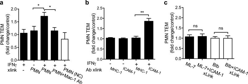
Figure 5. Engagement of apically expressed ICAM-1 induces MLCK-dependent increase in PMN TEM.
PMN TEM in the basolateral-to-apical direction across untreated (control) or IFNγ treated (to induce ICAM-1 expression) T84 monolayers, was induced by a gradient of fMLF (100nM). (A) Transmigrated PMN were collected and introduced to the apical side of new T84 monolayers in the presence of fMLF (100nM), or added to bottom chambers of transwells, facing the apical membrane, but without direct contact with the monolayers (2.5×10⁵ PMN/well, 1h). After washing off the apically adhered PMN, subsequent PMN TEM was quantified. PMN apical interactions specifically with IFNγ treated T84 IECs significantly increased PMN TEM. This effect was reversed in the presence of anti-Mac-1 inhibitory Ab (20μg/ml). For all conditions the number of PMN that remained adherent to the apical epithelial membrane after washes was determined and subtracted from the total number of transmigrated PMN. (B) PMN TEM after Ab-mediated crosslinking of ICAM-1 or control protein (MHC-1) was quantified. Crosslinking of ICAM-1 but not MHC-1 on IFNγ treated T84 cells significantly increased PMN TEM. (C) Control and IFNγ treated T84 monolayers were preincubated with ML- 7 (MLCK inhibitor, 20SM) or Blebbistatin (myosin motor II inhibitor, 10μm) alone or followed by ICAM-1 crosslinking. The effects of these inhibitors on ICAM-1 crosslinking induced increases in PMN TEM were quantified. Both inhibitors prevented ICAM-1 crosslinking induced increases in PMN TEM. For all panels the data presented as fold increase above PMN TEM across unstimulated T84 IECs. N=4 independent experiments in triplicates, *(p<0.05), **(p<0.01), n.s. not significant, ANOVA with Newman-Keuls multiple comparison test.

