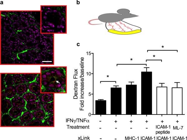
Figure 6. Engagement of ICAM-1 in murine intestine in-vivo leads to MLCK-dependent increase in intestinal permeability.
To induce ICAM-1 expression, mice were injected with a mixture of IFNγ and TNFα (500ng each, 24h, IP). (A) OCT-frozen segments of mouse intestine were sectioned (7μm-width) and immunofluorescently stained for ICAM-1 (green) and the tight junction protein, Claudin-2 (red). Representative confocal images show apical induction of ICAM-1 expression (white arrow) after IFNγ/TNFα treatment (bottom panel) compared to non-activated tissue (upper panel). The bar is 50μm. (B) Cartoon depicts the mouse intestinal loop model as described Methods. (C) In-vivo intestinal epithelial permeability was measured under the specified conditions as described in Methods. ICAM-1 tail peptide (100μm/ml) was introduced luminally 30 minutes prior to ICAM-1 crosslinking protocols. MLCK inhibitor ML-7 (20μM) was introduced by intraperitoneal injection 2.5 hours prior to ICAM-1 crosslinking. Increased FITC dextran flux after Ab-crosslinking of ICAM-1 was prevented with both the addition of ICAM-1 tail peptide and pretreatment with ML-7. N=4 mice/condition, *(p<0.05), ANOVA with Newman-Keuls multiple comparison test.

