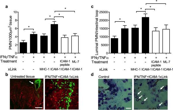
Figure 7. Engagement of ICAM-1 in murine intestine in-vivo leads to MLCK-dependent increase in PMN recruitment.
PMN TEM in the murine intestinal loop model was induced by luminal introduction of CXCL1 (1μM in 200μl HBSS+). (A) PMN in the mucosal epithelia were quantified from cryosections of the intestinal loop immunofluorescently labeled for PMN (green) and F-actin (red). (B) Representative images show robust PMN infiltration after ICAM-1 crosslinking (right panel) compared to intestinal tissue without introduction of CXCL1, where PMN are localized inside blood vessels (white arrow). The bar is 50μm. (C) PMN that migrated into the lumen were isolated by lavage and quantified from cytospins stained with Diff-Quik. Data presented as percent of all cells in the lavage fluid. (D) Representative images of the cytospins depict transmigrated PMN in the intestinal luminal lavage after ICAM-1 crosslinking (right panel, PMN are indicated by white arrows). PMN are not detected in the intestinal lumen without the introduction of the chemoattractant (left panel). The bar is 10μm. N=4 mice/condition, *(p<0.05), ANOVA with Newman-Keuls multiple comparison test.

