
Figure 1.
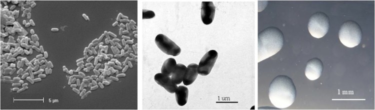
Images of Rhizobium leguminosarum bv. trifolii strain WSM2012 using scanning (Left) and transmission (Center) electron microscopy as well as light microscopy to visualize the colony morphology on a solid medium (Right).
Official websites use .gov
A
.gov website belongs to an official
government organization in the United States.
Secure .gov websites use HTTPS
A lock (
) or https:// means you've safely
connected to the .gov website. Share sensitive
information only on official, secure websites.
Images of Rhizobium leguminosarum bv. trifolii strain WSM2012 using scanning (Left) and transmission (Center) electron microscopy as well as light microscopy to visualize the colony morphology on a solid medium (Right).