Fig. 5.

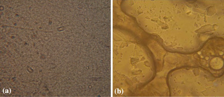
Light microscopy of nanoparticles produced with 1 % dextran concentration (A10). Freezing-point depressions to −20 resulted in disruption of nanoparticle configuration from normal (a) to sloppy crystalline shapes (b) (magnification ×400)

Light microscopy of nanoparticles produced with 1 % dextran concentration (A10). Freezing-point depressions to −20 resulted in disruption of nanoparticle configuration from normal (a) to sloppy crystalline shapes (b) (magnification ×400)