Figure 4.

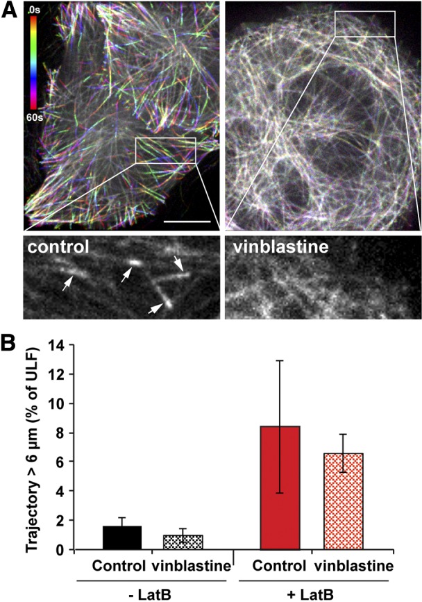
Microtubule dynamics is not required for ULF transport. GFP-ULF-expressing cells were transfected with TagRFP-EB3. Left panel; temporal color coding from the 60-frame projection of EB3 (1 frame/s) revealed the EB3 comet evolution at the tip of growing microtubules as an indicator of microtubule dynamics. Right panel; 10 nM vinblastine for 5 min is sufficient to stop microtubule dynamics, since EB3 comets at the microtubule tips are absent. Time color-code bar is shown in the control panel. Scale bar = 10 μm. Insets: enlargement of the first frame corresponding to the boxed area. Arrows point to the EB3 comets, which are present in the control cells. B) Vinblastine (10 nM) was added for 5 to 30 min to GFP-ULF cells treated or not with 5 μM Lat B. Graph shows the percentage of ULF tracks longer than 6 μm. Inhibition of microtubule dynamics does not substantially change the movement.
