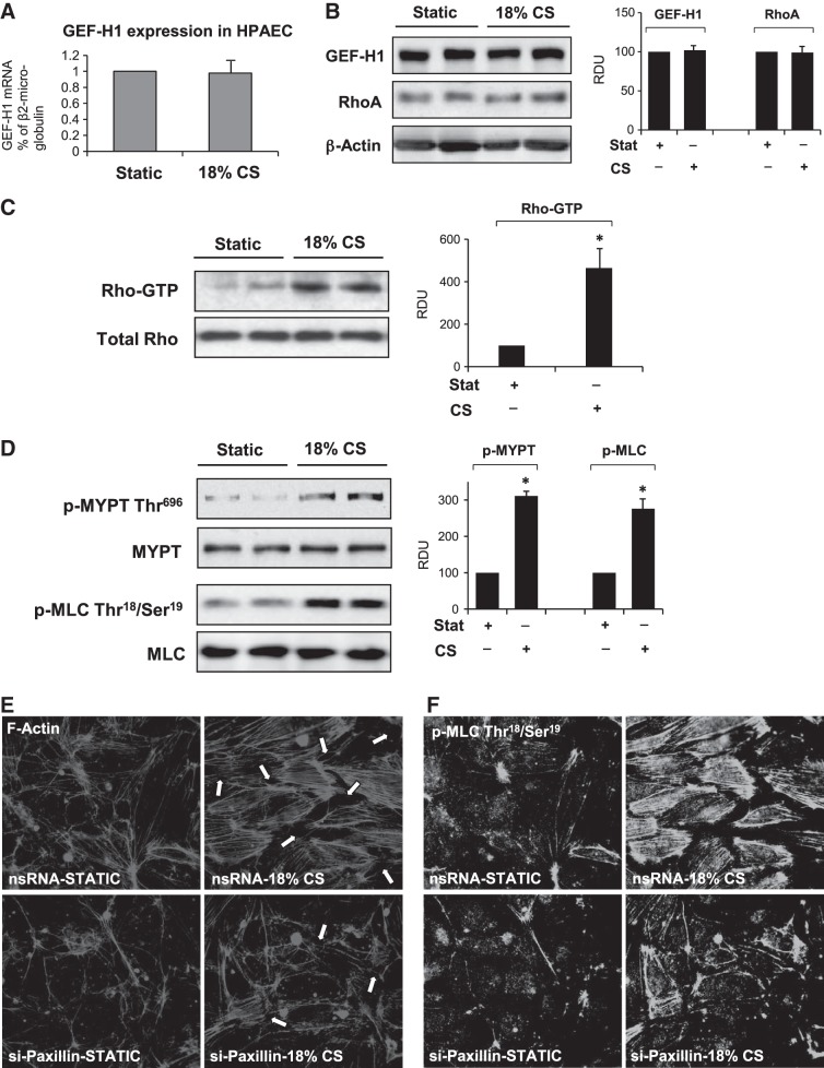
Figure 2.
Effect of acute CS on activation of Rho signaling. HPAECs were subjected to 18% CS for 15 min. A) GEF-H1 expression was analyzed by qRT-PCR. B) GEF-H1 and RhoA expression was analyzed by qRT-PCR Western blotting. C) CS-induced Rho activation was assessed by RhoGTP pulldown assay. D) Western blot analysis of CS-induced phosphorylation of MYPT and MLC. Probing for total protein was used as normalization control, n = 4 independent experiments. Bar graphs depict the quantitative densitometry analysis of Western blot data. *P < 0.05 vs. control. E, F) Human pulmonary ECs treated with paxillin-specific or nonspecific siRNA (nsRNA) were subjected to 18% CS for 15 min. E) F-actin remodeling in control and stretched EC monolayers was analyzed by immunofluorescence staining for F-actin. F) Intracellular localization of phosphorylated MLC was determined by immunofluorescence staining with phospho-MLC antibody.

