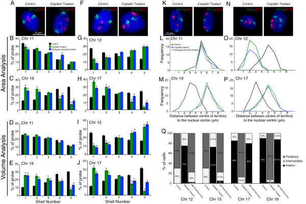
Figure 1.

Chromosome position in interphase nuclei of normal versus DNA-damaged fibroblasts. CT positions of chromosomes 11, 12, 17 and 19 as assessed by 2D-FISH and 3D-FISHin response to treatment with 25 μM cisplatin, 1 mM H2O2 and 0.05% dimethyl sulphoxide (DMSO) (control). (A, F) Representative images for 2D-FISH. (B, C, G, H) Signal intensity histograms of 100 nuclei divided into five shells of equal area. (D, E, I, J) Signal intensity histograms of 100 nuclei divided into five shells of equal volume. Error bars represent SEM. * indicates P = 0.05. (K, N) Three-dimensional projections of 3D-FISH. (L, M, O, P) Frequency distribution of distance (μm) between geometric centers of the CT and the nucleus for at least 50 nuclei per sample. (Q) Frequency distribution of cells with CTs (12, 15, 17 and 19) positioned in the nuclear interior, intermediate and periphery before and after damage (cisplatin treatment), and post cisplatin wash-off. For all datasets, the frequency distribution for cisplatin-treated samples was statistically significantly different from control and 24-hour post cisplatin wash-off samples (P = 0.001), while no statistical difference was observed between control and 24-hour post cisplatin wash-off samples.
