
Figure 2.
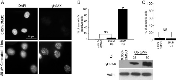
DNA damage caused by cisplatin treatment. Normal human dermal fibroblasts were treated with cisplatin and the extent of DNA damage and survival were monitored. Cells were treated for 4 hours with 25 μM cisplatin or 0.05% DMSO (control). γH2AX foci (A) and protein levels (D) increased in cisplatin-treated cells compared to their control counterparts. Annexin V staining (B) and FACS (C) were used to identify the percentage of cells undergoing apoptosis. ** indicates P = 0.001 NS: non-significant.