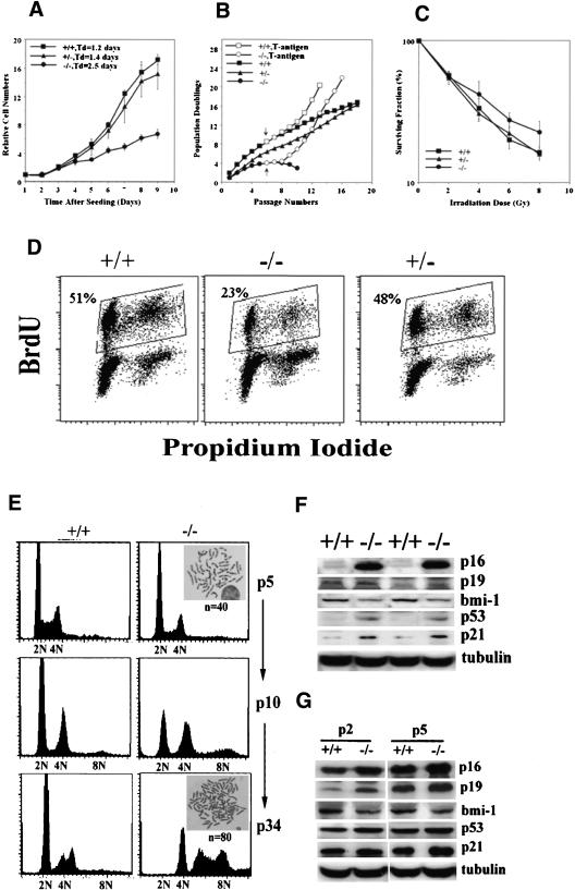
Figure 6.
PASG-/- phenotype is associated with decreased growth potential, replicative senescence, and altered expression of p16INK4a, p19ARF, bmi-1, p53, and p21. (A) Growth kinetics of MEFs (passage 1) demonstrate that the time to increase to fivefold the initial number of plated cells is 3.6, 3.8, and 6.0 d for wild-type, heterozygous, and homozygous mutant MEFs, respectively. The average of two cell lines of each genotype done in triplicate is shown. (B) Population doublings are lower in PASG-/- MEFs, which, however, can be rescued by SV40 Large T-antigen. The arrow indicates the time of transfection with SV40 large T-antigen containing retroviral supernatants. The average of two cell lines of each genotype done in triplicate is shown. (C) PASG-/- MEFs (passage 3) are more resistant to γ-irradiation than control MEFs. The number of surviving cells relative to unirradiated cultures of each genotype is plotted as a function of the dose of radiation. The average of two cell lines of each genotype done in triplicate is shown. (D) PASG-/--derived MEFs show a significant decrease of cycling cells. MEFs (passage 5) were continuously labeled with BrdU for 48 h and then analyzed by flow cytometry to determine the percentage of actively cycling cells. The average of two cell lines of each genotype is shown. (E) Aneuploidy is increased in PASG-/- MEFs over time. PASG-/- MEFs display diploid (n = 40) from early passages, but profoundly abnormal DNA content and increased chromosome numbers from late passages compared to comparatively stable wild-type MEFs. (F) Increased level of p16INK4a, p53, and p21 and decreased level of bmi-1 are observed in the kidney of PASG-/- mice. (G) Increased level of p16INK4a and p19ARF and decreased level of bmi-1 are observed in PASG-/- MEFs at passage 2 (P2) and passage 5 (P5).

