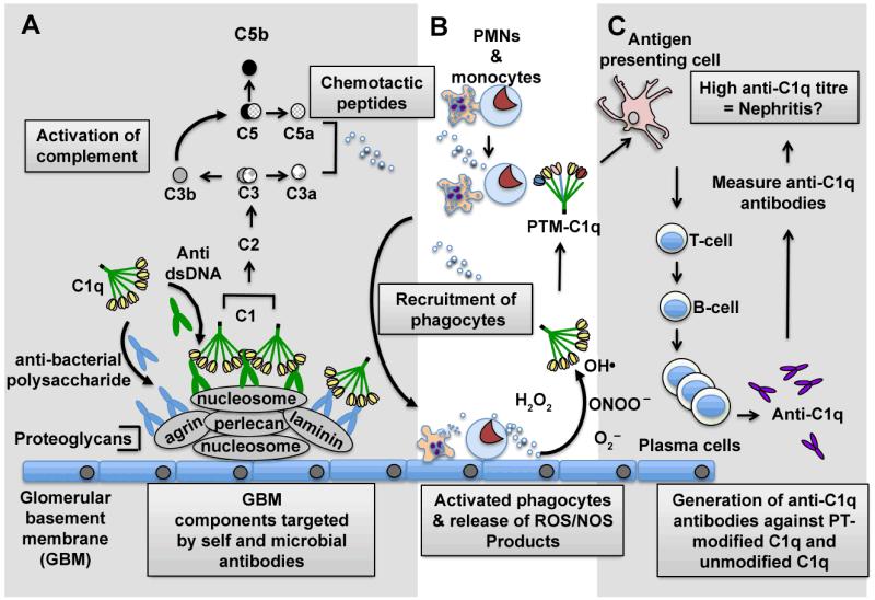
Figure 1. Postulated sequence of events in the generation of anti-C1q antibodies that may act as diagnostic biomarkers of glomerulonephritis.
Nucleosome blebs from apoptotic cells can deposit on the glomerular basement membrane (GBM) in SLE patients with LN and associate with a number of proteoglycan molecules. (A) During infection and/or inflammation anti-bacterial polysaccharide or anti-dsDNA antibodies bind to host proteoglycans and nucleosomes, respectively. This leads to the deposition of C1 (C1q/C1r2/C1s2) on the GBM and subsequent complement activation. (B) The release of chemotactic peptides C5a and C3a triggers the recruitment of phagocytes to the GBM in close proximity to C1q. The activation of the phagocytes leads to the release of free radicals that can post-translationally modify (PTM) C1q. (C) The PTM-C1q can be taken up by antigen presenting cells, and the modified peptides presented to T-cells. The autoreactive T-cells in turn can trigger B-cell activation and ultimately the production of anti-C1q-producing plasma cells. The concentration of anti-C1q antibodies produced in the blood can then detected by various immunoassays, including ELISA and used to assess whether a patient has or has not got nephritis.

