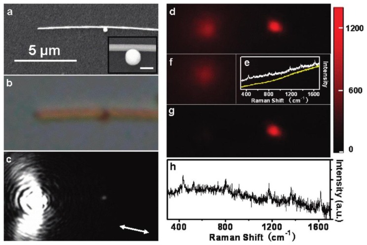
Figure 1.
Remote SERS of MGITC excited at the particle/nanowire junction. (a,b) Scanning electron microscope (SEM) and optical images of the structure, respectively; (c) Optical image of SPP propagation excited by 633 nm laser; (d) Raman image at 436 cm−1; (e) Spectra measured at the nanojunction (white) and the excitation spot (yellow); (f) Background fluorescence image of the substrate; (g,h) The corresponding Raman image and SERS spectrum after background correction. Adapted with permission from [49].

