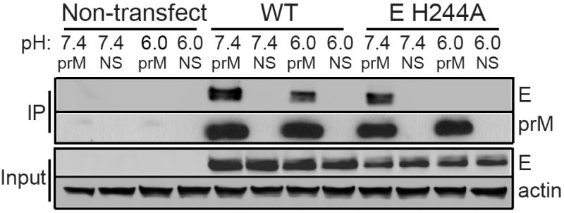
Figure 1. The M region of DENV prM protein binds E protein at neutral pH.
293T cells were transfected as indicated with plasmids encoding the WT or mutant prME (i.e. “E H244A”). Two days post-transfection, the cells were lysed in buffers at the indicated pH. Aliquots of the lysates were immunoprecipitated at the indicated pH with anti-prM mAb 38.1 (prM) or a non-specific mAb (NS), and analyzed by SDS-PAGE and western blot to detect prM or E. Expression levels in the lysates were evaluated by western blot analysis (input). The data are a representative example of 5 independent experiments. Full scans of the blots are in Supplementary Fig. 4.

