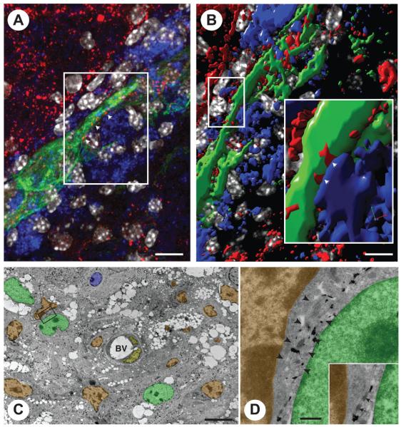
FIGURE 4.
NPC grafts establish juxtacrine signaling with endogenous professional phagocytes through junctional coupling. (A) Confocal microscopy image of GFP (green) NPCs contacting F4/80+ macrophages via connexin43+ cellular junctions (red; arrowheads). (B) Volocity V®-based 3D reconstruction of the confocal Z-stack in A. The magnified inset shows a structural junctional connexin43 pattern (red; arrowheads) between the process of one NPC (green) and one juxtaposed F4/80+ macrophage (blue). (C) Immunoelectron micrograph of GFP+ NPCs. The frame indicates one NPC whose processes are found to be in very close contact with a (GFP−) monocyte/macrophage. (D) High magnification of the frame in C showing the immunogold-labeled process of an NPC (arrowheads) running between a monocyte/macrophage and a second immunogold-labeled NPC. Cellular junctions between both NPC cytoplasms (inset, arrows) and between the NPC and the monocyte/macrophage can be observed in the inset. Pseudo colors in B and C: NPCs are in green; monocytes/macrophages are in orange; endothelial cells are in yellow and endogenous astrocytes are in blue. (Reproduced with permission from Cusimano et al., Brain, 2012, 135, 447-460, ©Oxford University Press.)

