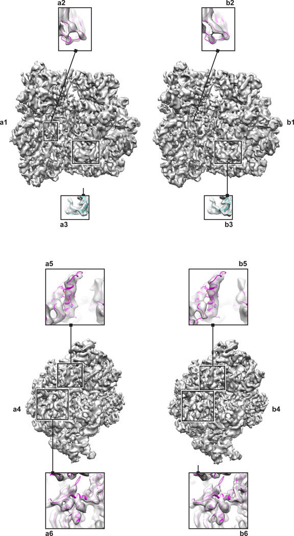
Figure 1.
70S Ribosome reconstructed from a) selections made by AutoPicker/ViCer b) manual verification performed by J.P. The left panel (a1, b1) shows the ribosome in the classic view with the 30S subunit on the left and the 50S on the right. Within this view, specific features highlight differences in resolvability of a loop (a2,b2) and a β-sheet (a3,b3). The right panel (a4,b4) shows an end on view of the 30S subunit.. Within this view, specific features highlight differences in resolvability of an α-helix (a5,b5) and the separation of secondary structure elements (a6,b6).

