
Figure 1.
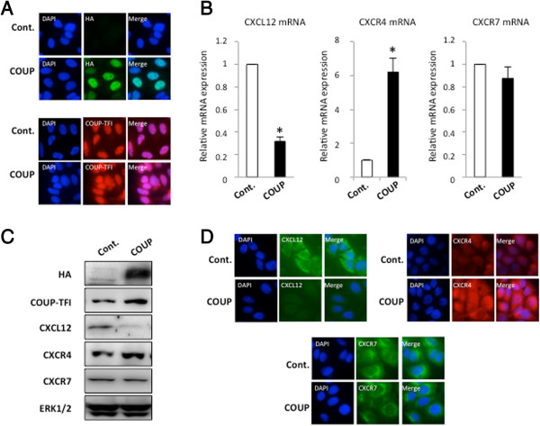
COUP-TFI modifies the expression of the CXCL12 signaling axis in MCF-7 cells. (A) Characterization of the control and COUP clones. An immunofluorescence cytochemistry assay was used to detect the relative expression of HA/COUP-TFI or COUP-TFI proteins in the control (Cont.) and COUP clones. The cells were fixed and processed for immunofluorescence as described in Methods; the nuclei were stained with DAPI. (B)CXCL12, CXCR4, and CXCR7 mRNAs were quantified by a real-time PCR analysis from two independent MCF-7 control and COUP clones. The results were normalized to GAPDH mRNA used as an internal control. The results were expressed as the relative mRNA expression level of CXCL12, CXCR4, or CXCR7. Data are the mean values ± SEM of at least three independent experiments. The asterisks indicate significant differences (p < 0.05) between the control and COUP clones. (C) The amount of intracellular HA/COUP-TFI, COUP-TFI, CXCL12, CXCR4, and CXCR7 protein was determined from whole-cell extracts of the different MCF-7 clones and compared to total ERK. A representative western blot is shown. (D) The control and COUP clones were fixed, and an immunofluorescence cytochemistry assay was used to detect the relative expression of CXCL12, CXCR4, and CXCR7 proteins. Staining with DAPI is also presented to visualize the nucleus of the cells.